Batrachospermum Occurrence:- (1) Batrachospermum is fresh water alga. (2) It is found in clear, cool, and running streams. (3) Deepwater plants are dark violet or reddish in color. But the shallow-water species are olive green. (4) The intensity of light changes the color of pigments. (5) The thallus is attached to the substratum. Vegetative structure (1) The thallus of an adult plant is soft, thick, filamentous. (2) It is freely branched and gelatinous. (3) The central axis is made up of a single row of large cells. Whorls of branches of limited growth are developed on this axis. (4) These branches are filamentous and dichotomously arranged. (5) The main axis is corticated. It consists of a row of elongated cylindrical cells....
Pinus
External
morphology :-
Root :-
The tree has a well developed tap root
system .
When the plant is growing on a shallow soil in the hills ,the lateral
roots spread very wide to fix the plant firmly into the plant firmly into
the soil.
 |
| Pinus Morphology |
Roots make association with
fungi in the soil called mycorrhizae.it is kind of symbiotic association where
the fungud does not affect the plant root bt provide soil nitrogen and phosphate
and other minerals from root to the root .
Stem :-
Stem is woodly and branched .the lower
unbranched part of the stem is called Trunk .
(1)
Branch of limited growth
(2)
Branch of unlimited growth .
The
branch is excurrent where the terminal bud or main stem shows more growth than
the terminal buds of lateral branches .lateral branches grow out from the main stem and are so
disposed that the plant gives a conical appearance .
Dwarf
Shoots are small a few mm. long and are
dot like .They arise in the axial of a scaly leaf on the long shoot .
Green leaves occur on the
dwarf shoots only .they also have scaly leaves
on them .
Leaves :-
Leaves are of two types :-
(a)
Foliage leaf :-
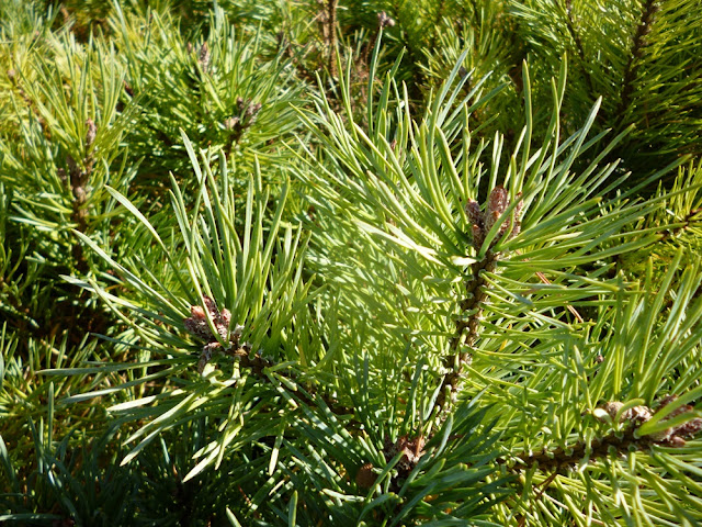
There leaves are long narrow ,green and look like
needles .
They occur in groups of 2-5 in different species on the tip of dwarf
shoots the dwarf shoot along with needles is also called a spur .
(b) Scaly leaves :-
They are small brown and membranous .
Scale leaves occur on
both long shoot as well as on dwarf shoots .
They are decious .However ,They are
protective and retain moisture around the stem.
Internal
Structure :-
T.S. Young Root:-
1. Outermost layer of the circular roots is thick-walled
epiblema with many root hair.
 |
| Young Root of Pinus |
2. Epiblema is followed by many layers of parenchymatous
cortex.
3. Inner to the cortex is present a layer of endodermis and
many layers of pericycle.
4. Vascular bundles are radially arranged and diarch to
tetrarch with exarch protoxylem.
5. Protoxylem is bifurcated (Y-shaped) towards the
periphery, and in between each bifurcation is present a resin cannal .
6. Phloem is present alternate to the protoxylem.
7. Pith is poorly-developed or absent.
T.S. Old Root Showing Secondary
Growth:-
1. On the outer side are present a few layers of cork,
formed by the meristematic activity of the cork cambium.
2. Cork cambium cuts secondary cortex towards inner side.
3. Many resin canals and stone cells are present in the
secondary cortex, the cells of which are separated with the intercellular
spaces.
4. Below the phloem patches develop cambium, which cuts
secondary phloem towards outer side and secondary xylem towards inner side.
5. Crushed primary phloem is present outside the secondary
phloem .
6. Many uniseriate medullary rays are present in the
secondary xylem.
7. Primary xylem is the same as in young roots, i.e., each
group is bifurcated (Y-shaped) and a resin canal is present in between the
bifurcation.
Long Shoot (Young):-
2.
Outermost single-layered, thick-walled
epidermis is heavily cuticularized and followed by multilayered cortex.
3.
A few outer layers of cortex are
sclerenchymatous, and some inner layers are parenchymatous.
4.
In the inner layers of
cortex are present many resin canals.
5.
The stele is eustelic or
polyfascicular endarch siphonostele.
6.
Vascular bundles are conjoint, collateral,
open and endarch, and resemble greatly with that of a dicot stem. 5-10 vascular
bundles are arranged in a ring.
7.
Endodermis and pericycle are indistinguishable.
8.
Narrow xylem rays connect the cortex and pith.
9.
Endarch xylem consists of only tracheids.
10. Phloem is
present on the ventral side and consists of sieve cells, sieve plates, phloem
parenchyma and some albuminous cells.
11. Intrafascicular cambium is present in
between the xylem and phloem.
12. Many leaf traces are also present.
13. A small parenchymatous pith is present in
the centre of stem.
T.S. Needle (Foliage
Leaf):-
1. It is
circular in outline in Pinus monophylla, semicircular in P. sylvestris and
triangular (Fig. 38) in P. longifolia, P. roxburghii, etc.
 |
| Foliage Leaf |
2. Outermost layer is
epidermis, which consists of thick-walled cells. It is covered by a very strong
cuticle.
3. Many sunken stomata are present on the epidermis.
4. Each stoma opens internally
into a substomatal cavity and externally into a respiratory cavity or
vestibule.
5. Below the epidermis are
present a few layers of thick-walled sclerenchymatous hypodermis. It is
well-developed at ridges.
6. In between the
hypodermis and endodermis is present the mesophyll tissue.
7. Cells of the mesophyll
are polygonal and filled with chloroplasts. Many peg-like infoldings of
cellulose also arise from the inner side of the wall of mesophyll cells.
8. Few resin canals are
present in the mesophyll, adjoining the hypodermis. Their number is variable
but generally they are two in number.
9. Endodermis is
single-layered with barrel-shaped cells and clear casparian strips.
10. Pericycle is
multilayered and consists of mainly parenchymatous cells and some
sclerenchymatous cells forming T-shaped girder, which separates two vascular
bundles .
Transfusion tissue consists of tracheidial cells.
Reproductive Structures of Pinus:-
1. Plant body is sporophytic.
2. Pinus is monoecious, and male and female flowers are
present in the form of cones or strobili on the separate branches of the same
plant.
3. Many male cones are present together in the form of
clusters, each of which consists of many microsporophylls. The female cones
consist of megasporophylls.
4. The male cones on the plant develop much earlier
than the female cones.
Male
Cone:-
Separate a male cone from the cluster, study its structure,
cut its longitudinal section, study the structure of a single microsporophyll,
and also prepare a slide of pollen grains and study.
2.
They replace the
dwarf shoots of the long shoot.
3.
Each male cone
is ovoid in shape and ranges from 1.5 to 2.5 cm. in length
4.
A male cone consists of a large number of
microsporophylls arranged spirally on the cone axis.
5.
Each microsporophyll is small, membranous,
brown-coloured structure.
6.
A
microsporophyll (Fig. 41) is comparable with the stamen of the flower of
angiosperms because it consists of a stalk (=filament) with a terminal leafy
expansion (= anther), the tip of which is projected upwards and called
apophysis.
7.
Two pouch-like microsporangia (= pollen sacs) are
present on the abaxial or undersurface of each microsporophyll. In each
microsporangium are present many microspores (= pollen grains).
8.
Each microspore or pollen grain is a rounded and
yellow-coloured, light, uninucleate structure with two outer coverings, i.e.,
thick outer exine and thin inner intine .
9.
The exine protrudes out on two sides in the form of two
balloon-shaped wings. Wings help in floating and dispersal of pollen grains.
10. Wings help in
floating and dispersal of pollen grains.
11. A few
microsporophylls of lower side of cone are sterile. Sporangia are also not
present on the adaxial surface of each microsporophyll of the male cone.
Female cone:
Observe the external features and longitudinal section
of a young female cone and also study 1st year, 2nd year and 3rd year female
cones.
1.
Female cone develops either solitary or in groups of 2
to 4.
2.
They also develop in the axil of scaly leaves on long
shoots like male cones.
3.
Each female cone is an ovoid, structure when young but
becomes elongated or cylindrical at maturity.
L.S. Female Cone:-
1. Many megasporophylls are arranged spirally on
the cone axis.
3. A few megasporophylls, present at the base and at
the apex of strobilus, are sterile.
4. Megasporophylls present in the middle of the strobilus
are very large and they decrease in size towards the base and apex.
 |
| Female cone |
5. Each megasporophyll consists of two types of scales,
known as bract scales and ovuliferous scales.
6. Bract scales are thin, dry, membranous, brown-
coloured structures having fringed upper part. These are also called carpellary
scales.
7. An ovuliferous scale is present on the upper surface
of each bract scale.
8. Each oruliferous scale is woody, bigger and stouter
than bract scale and it is triangular in shape. A broad sterile structure, with
pointed tip, is present at the apex of these scales. This is called apophysis.
9. At the base of upper surface of each ovuliferous
scale are present two sessile and naked ovules.
10. Micropyle of each ovule faces towards the cone
axis.
11. Each ovule is orthotropous, and it remains surrounded
by a single integument, consisting of an outer fleshy, a middle stony and an
inner fleshy layer. It opens with a mouth opening called micropyle.
12. Integument surrounds the megasporangium or
nucellus.
13. Just opposite the micropyle is present a pollen
chamber.
14. In the endosperm or female gametophyte are present
2 to 5 archegonia.
Female Cone of 1st Year :-
It is oval in
shape.
2. It
ranges from 1 to 4 cm. in length.
3. It is green to reddish-green in colour.
4. It is attached with the help of a short stalk on the
long shoot.
5. Megasporophylls are arranged very close to each
other, and so the cone is a compact structure.
Female
Cone of 2nd Year:-
1. It is elongated and larger than the first year cone.
2. It ranges from 5 to 15 cm. or more in length.
3. It is red-coloured structure.
4. It is woody in nature.
5. Megasporophylls are compactly arranged ,but not so
compact as in 1st year cone.
6. Seeds are present inside in the later stages
Female
Cone of 3rd Year :-
1. It is elongated or roughly rounded in shape.
2. It is also woody in nature like the 2nd year cone.
3. Megasporophylls are loosely arranged.
4. Seeds are dispersed from 3rd year cone.


Comments
Post a Comment
Thanks